Uterine fibroids non-cancerous tumor. The outer layer of tissue made of epithelial cells.

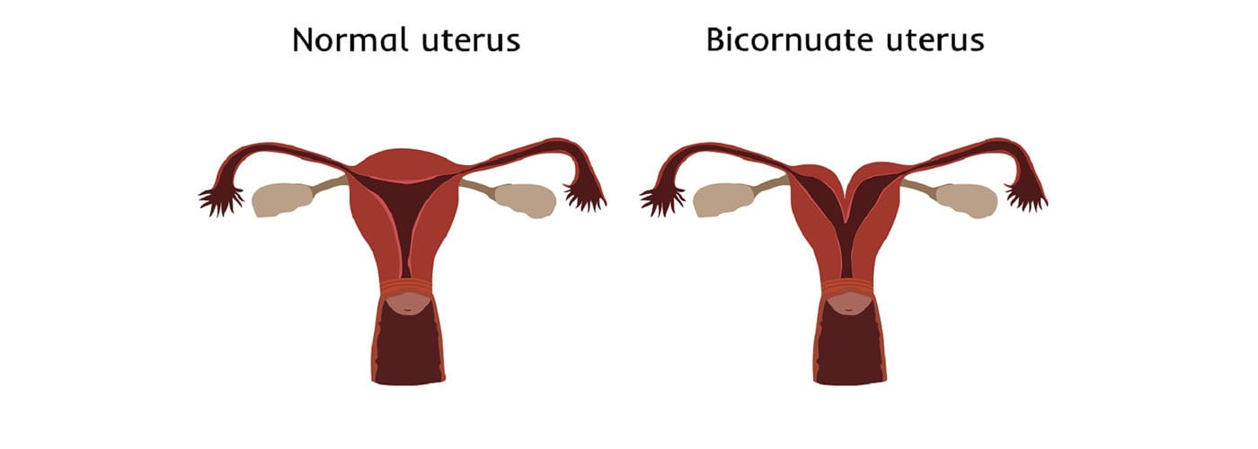
A bicornuate uterus is a heart-shaped uterusessentially a uterus with a dip on top.
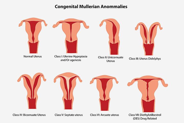
Different shaped uterus. A bicornuate uterus is a heart-shaped uterusessentially a uterus with a dip on top. Bicornuate uteri as well as unicornuate and didelphic uteri are considered mullerian duct abnormalities. A mullerian duct abnormality is a type of congenital abnormality of the uterus.
A heart-shaped uterus is actually a type of congenital uterine anomaly meaning that if you have one your uterus developed abnormally while you were still in the womb. If you have a bicornuate uterus it means that your uterus is heart-shaped. The uterus is the organ in a womans body that holds a baby.
This condition is sometimes referred to as a heart-shaped womb because it actually looks. The shape of your uterus is important if you become pregnant because it affects how a baby lies in your womb. Uterus irregularities are relatively unusual.
In a heart shaped uterus the two sides are joined at the lower end but the top has an indentation resulting in the characteristic heart shape. An irregularly-shaped uterus is relatively uncommon with only around 3 of women having any sort of abnormality in the shape size or formation of their uterus. Within this 3 a bicornuate uterus is the most common irregularity.
Normally symptom and trouble-free for. A heart-shaped uterus is actually a type of congenital uterine anomaly meaning if you have one your uterus developed abnormally while you were still in the womb says Megan Cheney MD an. Tilted uterus or retroflexed uterus symptoms include painful sex and the most pain when in the on-top sexual position along with painful periods.
Back pain discomfort when using a tampon and a predisposition to urinary tract infections are other signs that you might have a tipped uterus. A doctor should be able to tell you during a routine pap smear. Uterine fibroids non-cancerous tumor.
Uterus Types in Different Species Simplex No horns. Isthmus connected to uterine body Primates Duplex Large horns no body 2 cervices Rodents lagomorphs Bicornuate Has both uterine horns and uterine body pigs large horns. Cow mare sheep moderate horns and body.
The uterus has an inverted pear shape. In the adult it measures about 75 cm in length 5 cm wide at its upper part and nearly 25 cm in thickness. It weighs approximately 30-40 grams.
The uterus is divisible into two portions. Body and cervix. Generally speaking genetics and environmental factors determine the overall shape of your pelvis.
In the 1930s two researchers divided the pelvis into four different types. They largely based. The hysteroscope is introduced in the uterine cavity and the T shaped form is confirmed.
In case of using the Trophy hysteroscope gentle activation of the operative sheath is performed and a progressive visual dilatation of the cervical canal up to a diameter of 44 cm is performed Fig. Using the 5 Fr sharp scissors an incision line is made from the tubal ostium to the isthmus uteri. J David Steer P et al 2010 High risk pregnancy management options Elsevier Saunders Chan YY Jayaprakasan K et al 2011 Reproductive outcomes in women with congenital uterine anomalies.
A systematic reviewUltrasound Obstet Gynecol 38. RCOG 2011 Recurrent Miscarriage Investigation and Treatment of Couples Green-top Guideline 17 Royal. It is possible to identify three different subtypes of T-shaped uterus.
The most common type of T-shaped uterus with thick lateral walls and normal fundus without septum or subseptum appereance and interostial distance. The Y-shaped uterus with thick lateral walls fundal septum or subseptum and reduced interostial distance. And the I-shaped uterus with very thick lateral walls even above the.
Three distinct layers of tissue comprise the uterus. The outer layer of tissue made of epithelial cells. The middle layer made of smooth muscle tissue.
The inner lining that builds up over the course of a month and is shed if pregnancy does not occur. The womb uterus is shaped a bit like an upside-down pear tucked away in the pelvis. Its about 75cm long 45cm wide and 3cm deep.
Parmar et al 2016. If youve had a baby before your womb is likely to be slightly bigger than this. Parmar et al 2016 Verguts et al 2013.
The womb usually leans forwards towards the tummy. So even if you are diagnosed with a Heart-Shaped Uterus do not blame yourself the shape of the uterus isnt in your hands at all. In fact it is not even hereditary.
Also known as Bicornuate Uterus a Heart-shaped Uterus looks so due to its two paramesonephric ducts failing to fuse together. When not fused together the ducts give the appearance of a heart shape ie divided at the top. The uterus is 6 to 8 cm 24 to 31 inches long.
Its wall thickness is approximately 2 to 3 cm 08 to 12 inches. The width of the organ varies. It is generally about 6 cm wide at the fundus and only half this distance at the isthmus.
The uterine cavity opens into the vaginal cavity and the two make up what is commonly known as the birth canal.